Horse Anatomy
Anatomy and Physiology of Equine Joints
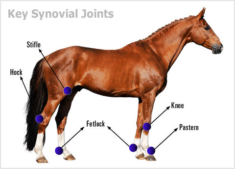
Joints allow the limbs to bend and the back to flex. Synovial joints are the joints of principal interest as they allow movement and are the type of joints between the vertebrae.
The synovial joint consists of two bone ends covered by articular cartilage. The articular cartilage is smooth and resilient and enables frictionless movement of the joint. The joint stability is maintained by a fibrous capsule which attaches to both bones and collateral ligaments. Collateral ligaments are important in maintaining stability in joints such as the fetlock, carpus, elbow, hock and stifle. In addition, there are other ligaments that also support the integrity of joints.
The joint capsule is made up of the fibrous capsule and an inner lining layer called the synovial membrane. The synovial membrane secretes the synovial fluid, which provides lubrication within the joint. There are various disease processes that affect the nature of the synovial fluid because of inflammation and disease in the synovial membrane. Inflammation in the joint causes excessive fluid production, which is due to synovitis (inflammation of the synovial membrane). The fluid produced by inflamed synovial membrane generally has a lower viscosity. This is a sign of disturbance in the production of hyaluronic acid, which is the key ingredient providing lubrication in the joint fluid.
Resilience of the cartilage tissue is important for normal motion as well as shock absorption. Hyaluronic acid provides lubrication to the synovial membrane surface and together with another protein, lubricin, it also lubricates the articular cartilage.
Limbs of the Horse
The limbs of the horse are structures made of many bones, joints, muscles, tendons and ligaments that support the weight of the horse’s body. The limbs play a major role in the movement of the horse, with the legs performing the functions of absorbing impact, bearing weight and providing thrust. In general, the majority of weight is borne by the front legs, while the rear legs provide propulsion. The hooves are also important structures, providing support, traction and shock absorption.
Good conformation in the limbs leads to improved movement and decreased likelihood of injuries.
Each forelimb of the horse runs from the scapula (shoulder blade) to the navicular bone (bone in the hoof). The bones and joints in between include:
- Humerus (arm)
- Radius (forearm)
- Ulna
- Elbow joint
- Carpus (knee) bone and joint
- Large metacarpal (cannon)
- Small metacarpal (splint)
- Fetlock joint
- Pastern joint
- Coffin joint

Each hind limb of the horse runs from the pelvis to the navicular bone. The bones and the joints in between include:
- Femur (thigh)
- Patella
- Stifle joint
- Tibia
- Fibula
- Tarsal (hock) bone and joint
- Large metacarpal
- Small metacarpal
- Fetlock joint
- Pastern joint
- Coffin joint

When the horse is moving, the coffin joint has the highest amount of stresses applied to any joint in the body, and it can be significantly affected by trimming and shoeing techniques. Although having a small range of movement, the pastern joint is also influential to the movement of the horse and can change the way that various shoeing techniques affect tendons and ligaments in the legs.
Joints in the Horse
Elbow Joint – The elbow joint is formed between the distal end (farthest) of the humerus and proximal ends (nearest) of the radius and ulna (which are fused in a horse). Flexion (bending) and extension are possible in the horse between the humerus and radius/ulna. The elbow is a typical synovial joint.

Stifle Joint – The stifle is the equivalent of the human knee and it is the largest, most complex joint in the horse. The bones that make up the stifle are the femur (thigh), tibia (shin) and patella (kneecap). The stifle lifts the leg upward and forward, making it critical to moving and athletic pursuits. The stifle has two joints, the femoropatellar joint (connects the kneecap) and the femororbital joint (connecting the bones). Stifle injuries can go undiagnosed because the stifle is difficult to evaluate. In addition to all common joint problems, traumatic injury to the bones and ligaments can occur due to activities involving sharp turns, while meniscal ligaments can be damaged by activities such as jumping. Stifle ligament and meniscal damage are currently the most under diagnosed.

Hip Joint – The horse has a limited range of hip movement compared to the dog. This is mainly restricted to bending and extension and is a result of conformation of its femoral head, intra-articular ligaments, and a large muscle mass around the joint. Disorders of the hip joint are relatively rare causes of lameness in horses. Most cases are traumatic in origin, secondary to falls, although septic disease and development disorders do occur.

Fetlock Joint – The fetlock joint occurs between the cannon bone, the proximal phalanx and sesamoid bones in the front legs. It allows bending and extension movements. The fetlock joint is arguably the joint that distinguishes a horse, with its unique anatomy and physiology allowing high speed, medium distance activity. The fetlock is a joint, a shock absorber, an energy storage system and a stabilizer of the front limbs. It is a complex joint with the bones and soft tissue interwoven. Development abnormalities and traumatic injury both shed debris into the joint which is the primary cause of joint disease. Degenerative arthritis is usually a secondary development.

Carpal Joint – The carpus (carpal joint) on a horse is commonly referred to as the “knee” which is only on the front legs. The tarsus is the corresponding joint on the hind leg, commonly called the “hock”. The horse’s knee is one of the most complex regions in the limb because there are several small bones and ligaments all combining to form the three main joints; the radiocarpal, intercarpal and carpometacarpal joints. There are 9 bones that make up these knee joints, two rows of small carpal bones join the radius at the top of the knee and the cannon and splints at the bottom. These bones are held together by a complex series of ligaments that help maintain stability, but also act as shock absorbers. Conformational defects are associated with an increased risk of injury and lameness. Osteoarthritis of the knee is by far the most common condition affecting this region in horses, and is often secondary to other problems such as chip fractures or poor conformation.

Tarsus Joint (Hock) – The hock is the joint between the tarsal bones and tibia. It is similar to the ankle of a human. Although the tarsus refers specifically to the bones and joints of the hock, most refer to the hock in such a way as to include the bones, joints and soft tissue of the area. The hock is especially important due to the great strain it receives, particularly when jumping and stopping. There are 4 joints within the hock, 3 of which permit almost no movement and are high impact, low motion joints. Because the hock takes a great deal of strain in all performance disciplines, correct conformation is essential if the horse is to have a sound and productive life.

Pastern Joint – The pastern joint is the joint between the long pastern bone (proximal phalanx) and the short pastern bone (middle phalanx). This is equivalent to the two largest bones in the human finger. This joint has limited movement but does help to disperse the concussive forces of the horse’s step and, also has some influence on the bending or extension of the entire leg. The pastern is vital in shock absorption, responsible for the concussive forces of a footfall, effectively helping to distribute the forces among the bones of the leg and the tendons and ligaments.

Coffin Joint – The coffin joint includes 3 bones, the middle phalanx (pastern bone), the distal phalanx (coffin bone) and the distal sesamoid (navicular bone). It allows slight bending and extension movements. The coffin bone is the wedge-shaped bone in the hoof that supports the horse’s weight. As 23-27% of the movement from the end of the cannon bone down to the ground is involved in the coffin joint, loss of appreciable movement in the coffin joint will impact the gait of the horse. Balanced shoeing and symmetry will maintain the coffin joint conformation and/or decrease the predisposition to coffin joint arthritis or inflammation.

Contact
DuoVital-Health LLC
P.O. Box 2115, Addison TX 75001
Tel: 1-469-291-5487
Mail: service@mobility-health.com
Blog
Company
Mobility Health News Room
Stay in the Know!